Hereditary Kidney Disease: The Genetics of PKD
PKD is a group of two genetic disorders that affect the kidneys and cause the formation of multiple fluid-filled cysts of various sizes. As these cysts grow, they squeeze and destroy normal kidney tissue, eventually leading to loss of kidney function. PKD is an inherited disease, meaning it’s passed down genetically from parent to child, just like eye or hair color. Polycystic kidney disease can be autosomal-dominant (ADPKD) affecting 50% of offspring. Or it can be autosomal-recessive (ARPKD), affecting 25% of offspring.
Mutations in one of two genes (PKD1 or PKD2) account for most cases of ADPKD. Polycystic kidney disease 1 gene (PKD1) mutations are the most common. About 80 percent of patients affected by ADPKD have a PKD1 mutation. PKD2 gene is the cause of up to 20% of ADPKD cases and it is usually associated with milder course.
The PKD1 and PKD2 genes provide the blueprints for important kidney and liver proteins called polycystin-1 (PC1) and polycystin-2 (PC2). These proteins are crucial for the structure of the kidney’s tubular cells, which filter and clean the blood. PC1 and PC2 influence healthy growth and fluid secretion in these cells. However, in people with hereditary kidney disease, abnormal genetic blueprints lead to the production of abnormal proteins. When these kidney proteins don’t work properly, cysts accumulate and damage the kidney.
Cysts that form in polycystic kidney disease usually occur when the cells lining the tubules of the kidney start growing out of control (called proliferation). These outgrowths bulge and eventually separate into cysts. As the cysts grow, they transport fluid across their lining, forming a fluid-filled sack much like a balloon. So, when we think about PKD, we should remember two processes: cell proliferation (cell growth) and fluid secretion into the cysts.
Truncating vs. Non-truncating mutations
Sometimes a change in the DNA sequence of a gene results in the creation of an “early stop.” This early stop functions to end the translation of the gene into a protein in our cell factory. This produces a shortened or truncated protein. This type of variant can have serious functional consequences. They are called truncating mutations.
The relationship between genetic variants and prognosis in PKD is not completely understood. In a study that looked at the “renal survival” in 741 patients with ADPKD, PKD2 mutations were associated with approximately 20 years longer survival than PKD1 mutations. In addition, the type of PKD1 mutation, not its position, correlated strongly with renal survival. The median age at onset of kidney failure was 55 years for carriers of a truncating mutation and 67 years for carriers of a non-truncating mutation. This observation allows the integration of genic and allelic effects into a single scheme, which may have prognostic value.
This points to the importance of genetic testing even in a genetic disease with an obvious clinical presentation such as PKD. Identifying patients with truncating mutations may help selecting those who require more aggressive therapies.
PKD at the Cellular Level
To determine the best PKD diet and lifestyle recommendations, we must understand what is happening at the cellular level. In essence, abnormalities in PC1 or PC2 proteins will activate two pathways inside the cell. First, it activates the cAMP pathway which regulates fluid transport. Second, it changes the way cells make energy from sugar (called glycolysis). When PC1 or PC2 proteins don’t work well, they switch from aerobic (with oxygen) to anaerobic (without oxygen) glycolysis. This is similar to the shift that occurs in cancer cells.
While anaerobic glycolysis produces less energy from glucose, it is faster than aerobic glycolysis. This faster energy production allows the cells to grow faster. However, it also leads to a critical dependence on glucose.1
In the next blog we will discuss the 2021 update about diet and Lifestyle Treatments to Improve Polycystic Kidney Disease (PKD).
1 It is noteworthy that glycolysis is inhibited by a cellular messenger called AMP-activated protein kinase (AMPK). It is activated by another messenger called mTOR. Metformin activates AMPK.
The post Polycystic Kidney Disease Genetics appeared first on Integrative Kidney.]]>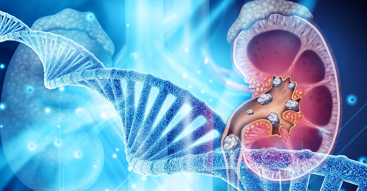
The gut-kidney connection refers to how a healthy gut barrier and diverse microbiome (the collection of friendly bacteria living in our intestines) directly influences kidney health. The connection between the gut and kidneys is very complex but we break it down into two major categories as we discussed in previous blogs: the gut-derived uremic toxins, and the inflammatory immune response that can trigger kidney disease.
In addition, as the gut is the gateway of nutrients into the body, it plays a major role in the balance of water and electrolytes such as sodium, potassium, magnesium and calcium.
The role of the gut in water and electrolyte balance
All of our water requirements are met by consuming food and beverages which are absorbed in the gut. In fact, water absorption in the gut is so powerful that, out of the 6 L of gastrointestinal secretions and 2 L of oral intake every day, only 150 ml are lost in the stool. Water absorption in the gut also depends on absorption of carbohydrates in the small intestine and short chain fatty acids (derived from the microbiome) in the large intestine.
Both the gut and the kidneys influence the body’s electrolyte balance. In most instances, electrolyte transport in the gut utilizes similar transporters to those found in the kidney tubules. Electrolytes are also regulated by hormones and neurotransmitters. These include neurotransmitters of the sympathetic system, compounds such as prostaglandins and hormones such as aldosterone.
Prostaglandins, for example, stimulate active electrolyte secretion in the small intestine and colon. Prostaglandins can also affect electrolyte handling by the kidneys. Aldosterone is well known to affect sodium, potassium and magnesium balance in the body by its effects on the kidneys and the colon.
The gut can also excrete water to help balance electrolytes in the body. Indeed, the gut is so effective in ridding the body of unwanted electrolytes that it can improve potassium balance in advanced stages of kidney disease.
The gut-kidney connection in electrolyte balance
Recently, a small protein called uroguanylin was discovered. It was found to regulate the transport of sodium in the gut and kidney and electrolyte balance by activating cGMP. Uroguanylin is often described as the “gut monitor of sodium intake.”
Most of what we know about uroguanylin so far comes from rat studies. For example, rats on low salt diets showed increased production of uroguanylin. If salt was given orally, there was a rapid increase in urinary salt excretion. However, if salt was given through the veins, urinary salt excretion was delayed. Similar findings were seen in humans but uroguanylin levels were not measured.
Other studies showed that giving intravenous uroguanylin can increase water, sodium and potassium excretion in the urine. These studies indicate that uroguanylin acts as a mediator of the gut-kidney axis because it regulates water and electrolyte balance.
The role of the gut in calcium balance
The gut also plays an important role in calcium balance. The strict balance of calcium in the blood is crucial for many biological functions. An interaction between the gut, the kidneys, and the bones helps maintain tight control over calcium levels.
Calcium is absorbed in the gut by two general mechanisms: transcellular active transport process and a passive paracellular (in between the cells) process.
Active transport mainly occurs in the first part of the gut (duodenum and upper jejunum). It plays an important role in absorbing calcium when calcium intake is too low. In active transport, calcium enters the cells of the gut through calcium-selective ion channels (CaT1). Calcium is then transported within the cell by calcium-binding protein (CaBP) and it is deposited into the bloodstream by a calcium ATPase pump (CaATPase).
Passive transport occurs throughout the gut. It is most important when calcium intake is high. Paracellular transport of calcium depends on the difference in electricity and concentration across the cells in the gut lining. It involves specialized proteins called claudins.
The table describes key differences between the two mechanisms of calcium absorption in the gut.
| Calcium Absorption in the Gut | ||
| Transcellular | Paracellular | |
| Energy required | Active | Passive |
| Location | Upper small intestine | Throughout the intestine |
| Transporters involved | CaT1, CaBP, CaATPase | Claudins |
| Baseline dietary status | Low calcium intake | High calcium intake |
Like the gut, the kidneys are responsible for maintaining calcium balance. The kidney tubules recycle needed calcium and add it back to the blood, a process called reabsorption. Unlike calcium transport in the gut, most calcium reabsorption in the kidneys occurs via a passive paracellular transport in the proximal tubules. When the kidneys filter excess calcium out of the blood, calcium is excreted in the urine through active transcellular transport in the distal tubules. Active transport utilizes calcium channels while passive transport also utilizes claudins.
Claudins
Claudins are a family of proteins that participate in forming strong and healthy barriers between cells that line the gut and the kidneys. These barriers are important to prevent “leaking” in between cells and to keep bad things out of the bloodstream. The space between two cells that fit closely together is called a tight junction. Claudin-2 is found in the tight junctions of the proximal tubule of the kidney and plays an important role in paracellular calcium reabsorption. What is fascinating is that claudin-2 was also found to be part of the tight junctions in the gut. Studies have demonstrated that claudin-2 is a mediator of intestinal permeability during intestinal inflammation. Read more about intestinal permeability here.
Genetic variation in claudin-2
Genetic variations in the gene that codes for claudin-2 (CLDN2) have been documented to decrease the paracellular “leak” of calcium. So genetic variations in the CLDN2 make the tight junctions tighter and the barrier stronger. This change decreases the ability of the kidneys to filter calcium and add it back to the blood. Since calcium cannot be properly recycled, it is excreted in the urine at high levels and increases the risk for kidney stones.
Interestingly, the increase in calcium in the urine is not accompanied by an increase in blood calcium, parathyroid hormone, or 1,25 dihydroxy vitamin D3 levels. This indicates that there is an accompanied increase in absorption of calcium in the gut. This will help maintain calcium balance in the blood which is crucial for many biological functions.
In essence, the same genetic variations in CLDN2 that lead to increased calcium in the urine also lead to an increase in calcium absorption in the gut. This effectively maintains calcium balance in the blood but increases the risk for kidney stones. This was proven in a study showing that low calcium in the diet leads to low calcium in the urine of people with the CLDN2 genetic variation.
The bottom line
There is also a gut-kidney connection when it comes to electrolyte balances in the blood. Understanding the mechanism and the genetic basis of electrolyte disorders may help individualize treatment for kidney patients. Kidney stone patients with CLDN2 genetic variants, for example, may benefit from dietary calcium restrictions which is generally not recommended for all kidney stone patients. We utilize Natera’s Renasight broad panel genetic test to identify the genetic risk for kidney stones. For more information, check out our blog about the integrative approach to kidney stones.
The post Genetics of the Gut-Kidney Connection in Kidney Stones – Beyond the Microbiome appeared first on Integrative Kidney.]]>

Genetic testing for patients with kidney disease can have a remarkable impact on their care. The availability of “broad-panel genetic testing” for kidney patients ushers in a new era of nephrology and patient care. Tests that used to cost thousands of dollars and took months for results can now be done for a fraction of the cost in just a few weeks. New commercially available genetic tests utilize next-generation sequencing to identify multiple gene variants simultaneously. These tests can help in the management of kidney disease in multiple ways. In this blog, we will focus on the clinical utility of genetic testing for kidney transplant evaluation.
Genetic Testing for Kidney Transplant Evaluation
During the evaluation of a patient for a kidney transplant (the recipient), the assessment usually focuses on answering these questions:
- Can the recipient survive elective transplant surgery?
- Can the recipient tolerate immunosuppression after the transplant?
- Can the recipient have a good outcome?
In addition, evaluation of living donors try to answer questions about their suitability for donation and their risk of developing kidney failure in the future.
Living Donor Evaluation
One of the most pressing questions when evaluating a living donor is: Will this donor develop kidney disease in the future if s/he donates a kidney now? Several studies have shown an increased risk of the donor developing kidney disease after donation. This risk is higher if the donor and the recipient are related. This may indicate that genetic factors play a role in this risk.
In 2017, the Kidney Disease Improving Global Outcomes (KDIGO) Guidelines suggested that “transplant programs should have a strategy for evaluating for inherited kidney disease in donor candidates when there is a family history of kidney failure and the recipient’s cause of kidney failure is uncertain.”
These guidelines suggested genetic testing of living related donors with specific diseases such as focal segmental glomerulosclerosis (FSGS), atypical hemolytic uremic syndrome, Alport disease, sickle cell trait, and autosomal dominant tubulointerstitial kidney disease.
Genetic testing of a living relative donor can be especially important if the recipient has polycystic kidney disease. If this mutation is identified in the recipient, the donor can then be tested and excluded if s/he has the mutation. This can give greater assurance to both the donor and recipient.
Other genetic variants are associated with increased risk of chronic kidney disease (CKD) such as APOL1 gene variants that are associated with increased risk for nephropathy in patients of African ancestry. Incorporating testing for these genetic risk variants in the evaluation of the donor may help replace race for calculation of the so-called Kidney Donor Risk Index that is used to predict the longevity of the transplant graft.
While it is still too early to incorporate the genetic risk variants for diabetic kidney disease and IgA nephropathy in transplant evaluation, getting more clarity on the utility of the risk variants can have a tremendous impact on the care of current patients.
Recipient Evaluation
Kidney disease is silent in its progression and symptoms do not develop until the advanced stages of CKD. One in 10 patients with advanced kidney diseases presents with end-stage kidney disease (ESKD). In many of these cases, the laboratory workup is inconclusive, and their kidneys are often too atrophic to biopsy. Unlike kidney biopsies, genetic data can be informative even after ESKD has developed.
Genetic evaluation of the recipient is, therefore, helpful in identifying the causative mutation that could have led to the disease. Using targeted gene testing, researchers were able to identify pathogenic mutations in 19% of waitlisted transplant patients under the age of 40. Broad panel genetic testing can likely have an even higher yield. Indeed, broad panel genetic testing has been shown to identify the cause of CKD in up to one-third of the patients with an unknown cause.
Genetic testing of the recipient can also help in providing individualized post-transplant care. Finding a specific mutation that leads to a localized disease in the kidneys can decrease concerns about the recurrence of the disease after transplantation.
Also, a genetic diagnosis can often point to the likelihood of disease in another organ and can prompt referral and evaluation.
Currently, researchers are collecting phenotypic and genetic information on patients receiving transplants in the iGeneTRAiN consortium. Analyzing this data in the future may have a significant impact on our understanding of transplant graft outcome.
Pharmacogenomics
Wouldn’t it be a relief to be able to predict in advance how someone might respond to a medication? This would save time, eliminate guesswork, and improve patient outcomes. Thanks to advances in a field of genetics called pharmacogenomics (PGx), clinicians have begun to use genetic information to personalize drug therapy.
Accurate pharmacogenomics data are now available on two transplant medications: tacrolimus and azathioprine. Although the latter is not commonly used, the former is used often. Tacrolimus is metabolized by the enzyme encoded in the gene CYP3A5. Variants in this gene can classify the patient into one of three phenotypes: extensive metabolizer, intermediate metabolizer, and poor metabolizer. Indeed, pharmacogenomic data can now be used to optimize the initial dose of tacrolimus.
Many other medications commonly used by patients have pharmacogenomic data which can also be used to optimize their dosing. Medications such as clopidogrel, voriconazole, and allopurinol are a few of these. We discussed these medications in-depth in our previous blog about pharmacogenomics.
The Bottom Line
Genetic testing is gradually becoming a significant part of the transplant evaluation of the donor and the recipient. It is particularly useful in the evaluation of living donors with a family history of kidney disease. This data has the potential to transform the care of kidney transplant patients and improve their outcomes.
The post Clinical Utility of Genetic Testing for Kidney Transplant Evaluation appeared first on Integrative Kidney.]]>
FAQs about genetic testing for kidney disease
What can I learn from genetic testing?
Genetic testing for kidney disease can help diagnose unknown causes of chronic kidney disease (CKD) and help identify changes in the genome that may increase CKD risk. Results from the tests can even help providers correct improper diagnoses. In addition, genetic testing can determine kidney disease severity. Identifying specific gene variants can help guide treatment for specific types of CKD as well as proper dosing of medications.
How does it work?
In order to perform the test, a sample of DNA is required, usually obtained through a saliva or blood sample. The cells from the sample are processed and analyzed in a database, obtained from Genome Wide Association studies (GWAS), of currently known genetic variants associated with CKD. These studies looked at multiple genetic variants and linked them statistically to specific kidney diseases in established patients.
Does genetic testing help diagnose specific kidney disease?
As mentioned above, genetic testing can help identify genetic variants associated with certain kidney diseases. However, carrying a genetic variant does not necessarily mean a person will develop the disease. Other factors can play a role in the genes’ ability to express themselves. Genetic tests should always be interpreted in consultation with an experienced nephrologist or genetic counselor.
Will these tests identify all genetic variants associated with CKD?
No, our knowledge about the genetic basis for CKD is still evolving. To date, mutations in more than 500 genes have been associated with different forms of kidney disease. The specific genetic mutations identified on the panel varies depending on the genetic testing company used. In addition, genetic testing cannot tell you everything about inherited diseases. Diet, lifestyle, and environment all influence how genes are expressed, a field known as epigenetics.
Why do I need genetic testing for kidney disease if I am already doing regular physicals and lab checks?
Kidney disease is a silent disease and patients usually don’t develop symptoms until later stages of CKD. In addition to laboratory testing, genetic testing provides information for those at increased risk of CKD. Knowing your genetic risk provides you with an opportunity to modify your lifestyle in order to decrease the chance of kidney disease and failure in the future.
Is this test covered by my health insurance?
Depending on the test performed, your insurance may or may not cover a portion of the cost. Some small panel tests implemented by a few pharmaceutical companies are free. These companies hope to recoup the cost by identifying patients who will benefit from their products. In general, broad panel genetic tests are now much more affordable than they used to be.
Will I be discriminated against based on my results?
Federal law prohibits health insurance providers and employers from discriminating against a person based on genetic information. However, unfortunately, this law does not apply to long-term care, disability, or life insurance providers. It is crucial that you choose a testing company that values your privacy.
The Bottom Line
Genetics are one factor that plays a significant role in the development of kidney disease. Genome-wide association studies (GWAS) have identified several hundred genes that are associated with kidney diseases. Therefore, genetic testing may play a crucial role in the management of kidney disease patients.
The post FAQs About Genetic Testing for Kidney Disease appeared first on Integrative Kidney.]]>
Current evidence suggests that genetics play a role in the development of kidney disease. Common genetic disorders associated with kidney disease include polycystic kidney disease, Alport’s Syndrome and Fabry’s disease. The advances of genome-wide association studies (GWAS) helped identify several hundred other genes linked to kidney diseases. This made genetic testing a useful tool in the management of kidney disease patients. In this blog, we will detail the benefits of broad-panel genetic testing in kidney disease management.
Next Generation Sequencing
Various methods for identifying genetic variants have been used in the past. The exome is that 2% of the genome that codes for all biological proteins. Next generation sequencing is a new technology that allows DNA sequencing of the entire human exome within a single day. It also captures a broader spectrum of variations that can affect the genetic code. This revolutionary technology can identify mutations associated with CKD. It can also recognize variants associated with an increase in risk or severity of CKD. It is now available for kidney disease patients. This type of “broad-panel genetic testing” can transform care for patients with kidney disease.
Clinical benefits of genetic testing in kidney disease management
Identify diagnosis of unknown causes
While diabetic and hypertensive kidney diseases are the most common causes of kidney failure, there are others. There are instances when providers find themselves unable to identify the cause of kidney disease. Indeed, in some cases, urinalysis is actually “bland” and the workup to identify the cause of kidney disease is negative.
Even a kidney biopsy may not be helpful. It may show glomerulosclerosis and fibrosis (scarring). This does not help identify the original cause of kidney disease. Genetic testing can have a tremendous value in these cases. In fact, whole exome sequencing was able to diagnose up to one third of the patients who had unknown causes of kidney disease.
Reclassify a clinical diagnosis
It is common for providers to label kidney disease patients with unknown causes as hypertensive kidney disease or “nephrosclerosis”. This is because hypertension is common in kidney disease patients. Studies have shown that 60-90% of patients with chronic kidney diseases have high blood pressure. It is often not clear which came first, hypertension or kidney disease. It is possible that patients who were diagnosed with hypertensive kidney disease have another cause. In fact, studies have shown that up to a quarter of kidney diseases can be reclassified with a broad-panel genetic testing.
Target the therapy and workup
Broad-panel genetic testing can help providers avoid unnecessary procedures, tests, and treatments. It gives accurate and “molecular level diagnosis”. It provides a better idea of the outcome of the specific kidney disease. It also helps providers avoid the use of immunosuppressive medications in patients with genetic causes of kidney disease. Furthermore, it can guide therapy for specific genetic causes that we currently have treatment for such as Fabry’s disease. This type of testing may, indeed, eliminate the need for a kidney biopsy.
Role in kidney transplant
Genetic testing can have a tremendous impact in guiding kidney transplant and in the care of kidney transplant recipients. Kidney transplant donors can be pre-screened by broad panel genetic testing to assess if they carry any genetic kidney disease risk. While carrying the genes does not necessarily indicate that the donor will have the disease, it can play an important role in selection. This is especially true for living donor kidney transplant when the recipient has a known genetic kidney disease, and the donor is too young to have any manifestations.
In addition, many immunosuppressive medications that are used by kidney transplant patients are metabolized by well-established pathways that can be affected by genetic SNPs.
Having this genetic information can help providers prescribe the proper dose of immunosuppressive medications which is critical in transplant patients to avoid rejection or toxicity. This evolving field is called pharmacogenomics.
Help with management of kidney patients
Broad panel genetic testing can identify patients who are at risk for kidney disease, reclassify the exact cause of kidney disease, and guide treatment. Knowing the molecular basis of some kidney diseases can guide lifestyle modification interventions, future development of drugs, and gene therapy. It can also identify early complications outside the kidneys that can be related to the specific genetic disease and prompt early interventions.
Available broad panel genetic tests
Broad panel genetic tests are now available for providers and patients, and are relatively inexpensive. Many of them are covered by insurance companies. We have utilized Natera’s Renasight broad panel genetic test for this purpose. It provides next generation sequencing for 382 genes that are associated with kidney disease. There are also other genetic testing companies that test a small panel of genes for free to identify patients for drug or gene therapy.
The Bottom Line
We are at the dawn of a new era in nephrology and kidney care. Broad panel genetic testing will revolutionize kidney disease management. Our genes are not our destiny, but we cannot change our destiny without knowing our genes.
The post Genetic testing in kidney disease appeared first on Integrative Kidney.]]>
The basics
As you may remember, each person inherits 46 chromosomes, 23 from each parent. These chromosomes house the genetic code that determine the characteristics of each person. They are composed of DNA which is a long and windy spiral made up of millions of nucleotides. There are 4 nucleotide bases (adenine, cytosine, guanine, thymine), the sequence of which determines how genetic traits are expressed.
This nucleotide sequence forms the foundation of about 22,000 genes. Our cells read the genetic sequence and use it to make thousands of proteins which are essential for carrying out biological functions that maintain life. In other words, you can think of the cells as protein factories. The DNA code provides the blueprint for all the workers to create proteins. These proteins can function as enzymes, receptors, or other structures that are important for sustaining life.
Errors in the code
Alterations in the sequence of nucleotide bases cause errors in the code and affect the efficiency of the protein production process. These may sometimes lead to disease or increased risk for disease. Interestingly, on occasion they may have no effect on risk and may even be protective against disease. There are three major changes that can occur in the code:
- Single-nucleotide substitution: this is also called single-nucleotide polymorphism or SNPs. These are common.
- Insertion or deletions (indels): a small stretch of DNA can be inserted or deleted from the code.
- Structural variation: a large-scale change or rearrangement in the DNA.
It is fascinating that a single variation in one nucleotide among the set of 3 billion in our DNA can sometimes cause a severe and deadly disease and other times have no effect whatsoever.
Why genetics are important?
Genetics play an essential role in many functions in the human body. Genetic changes can affect the type of food that a person prefers to eat. For example, genetic variants in bitter taste receptors in the tongue have been associated with decreased intake of vegetables and increased obesity. Genetic changes can also affect digestion, and absorption of food. They can modify the way we metabolize drugs, and toxins. They can alter the function of vitamins and other nutrients and their interactions.
The genetics of disease risk
For a long time, scientists have been trying to study how variations in genes lead to diseases. Past efforts focused on identifying inherited diseases in the so-called Mendelian patterns. However, recent advances in GWAS studies are making it more possible to identify genetic changes that can be associated with increased risk of a disease. Genetic variations in one or several genes can lead to minor changes that collectively may increase the susceptibility for certain common diseases such as diabetes and high blood pressure. This is an evolving field of study and we will continue to learn about it every day.
Genetics of kidney disease
There are well known and relatively common genetic disorders such polycystic kidney disease, Alport’s Syndrome and Fabry’s disease. GWAS studies has identified more than 500 genes that are associated with kidney diseases. Current evidence suggests that a significant genetic component plays a role in the development of kidney disease. These are evident from the ethnic variabilities in certain diseases such as the higher incidence of IgA nephropathy among Asians.
Genetics can also determine the severity of a kidney disease, the age of onset and the likelihood of ending up with End-Stage Kidney Disease. For example, different gene mutations that can cause polycystic kidney disease can have different outcomes.
In addition, genetic variations can explain the differences in susceptibility of the kidneys to systemic disease such as diabetes and lupus. These variations may explain why some people with diabetes get severe kidney disease while others don’t.
Finally, alterations in several genes have been associated with increased risk for CKD. There are several candidate genes. Mutations in the UMOD that encodes uromodulin is one of these candidates. Uromodulin, which used to be called Tamm-Horsfall protein, is excreted in certain portions of tubules and protects from urinary tract infections. The Shroom3 gene is another candidate that has been associated with increased risk for CKD.
Next generation sequencing
Various methods for identifying genetic variants have been used in the past. Next generation sequencing is a state-of-the-art technology that allows DNA sequencing of the entire human genome within a single day. It also captures a broader spectrum of variations that can affect the genetic code. This revolutionary technology is now available for kidney disease patients and can identify various mutations that are associated with CKD or can increase the risk or severity of CKD.
The Bottom Line
Genetics are one of the factors that lead to the development of kidney disease. For genes and their variants to contribute to a disease, we need to understand all of the factors that explain why people with the same variant may have different outcomes. Understanding the interaction between genetics, epigenetics and environment are crucial in this regard. Ongoing research in this field will not only increase our understanding of kidney disease risk but our ability to find various lifestyle modifications and therapies that can help decrease the burden of CKD worldwide.
Professionals: You can get free CME and learn more about genetics of kidney disease here.
The post Genetics of Chronic Kidney Disease appeared first on Integrative Kidney.]]>In this series we’re focusing on the integrative approach to preventing kidney stone formation.
Conventional approaches to kidney stones tend to focus on medications, surgical removal, and using ultrasonic waves to break up stone. It rarely approaches the root cause including risk factors to prevent stone formation.
We covered the impact of diet, the microbiome and gut health, and electrolyte imbalances on kidney stones in previous blogs. Here we will discuss the role genetics play in kidney stone formation.
The Genetics of Kidney Stones
There seems to be a familial link when it comes to the development of kidney stones. In fact, two thirds of patients with calcium-containing kidney stones have a relative with kidney stones. Recently, genome-wide association studies uncovered several genetic sequence variants (SNPs) that lead to increased risk of kidney stone development. Although we are still scratching the surface in understanding the contribution of genetic factors to stone formation, we do know that we can modulate these risks through environmental and dietary modifications.
Genetic Variations in Calcium Handling
In a previous blog, we discussed the role of the kidney filtration units (specifically the nephrons). The kidneys are responsible for filtering large volumes of blood daily. This function is crucial, and, the unique design of the nephrons make it able to adjust this filtrate and prevent dehydration. This intricate design also makes the kidneys crucial in the balance of water and many electrolytes.
Calcium is one of the electrolytes filtered and reabsorbed in the kidneys. Calcium sensing receptors (CaSR) are present in kidney cells and are essential for the reabsorption of calcium. These receptors increase and decrease the amount of calcium reabsorbed based on the calcium level in the blood. In other words, activating these receptors increases the amount of calcium lost in the urine.
Single nucleotide polymorphisms (SNPs) in CaSR have been found to alter the function of these receptors leading to increased urinary excretion of calcium. Of particular interest, SNPs in rs7652589 and rs1501899 were associated with kidney stones in patients with normal citrate excretion. SNPS in rs1801725, rs1042636 of the CaSR gene were also associated with kidney stones in various populations.
Another gene that has been associated with increased calcium in the urine is the gene coding for the protein Claudin-14, responsible for forming tight junctions. This protein helps connect adjacent cells to form a barrier which acts like a gate, separating blood from urine. Tight junctions are also responsible for ensuring that minerals don’t pass between the cells. SNPs in the genes that code Claudin-14 (CLDN14) alters the integrity of these “gates” and allows for calcium to “sneak” between cells into the urine, increasing the risk for kidney stone formation. Specifically, SNPs at the locations rs219778, and rs219780 of the CLDN14 gene were significantly associated with kidney stones.
Genetic Variations in Vitamin D Receptors (VDR)
Vitamin D plays a crucial role in calcium balance. Studies have shown that vitamin D increases the absorption of calcium from the gut and also increases calcium excretion in the urine. Vitamin D receptors are essential for vitamin D to exert its action on calcium balance.
Some mutations or SNPs in the VDR are associated with increased absorption and excretion of calcium, significantly increasing the risk of kidney stones. It is worth mentioning here that there is some controversy about the link between vitamin D supplementation and kidney stone formation. Vitamin D deficiency appears to be common among kidney stone formers. This is likely because low vitamin D causes calcium loss from the bone in order to maintain normal calcium range in the blood for cardiovascular protection. Even though vitamin D3 supplementation may increase calcium excretion in the urine, it has not been conclusively found to increase the risk of kidney stone formation.
Therefore, genetic assessment may be a key to identify patients who are at risk of kidney stone formation from taking vitamin D supplements.
Genetic Variations in the Handling of Other Minerals
The kidneys are crucial for balancing many minerals in our bodies such as magnesium, phosphate, oxalate and others. Genetic mutation or SNPs affecting the genes that code for the channels or receptors that regulate these minerals can also impact the risk of kidney stones. Some SNPs on the other hand can be protective against kidney stones such as SNPs in the UMOD gene. Discussion of this long list of SNPs requires details beyond the scope of this blog, but we summarized most of the genes that have been associated with kidney stones in the table below.
| Gene symbol | Gene name | Phenotype |
| ADCT10 | Adenylate cyclase 10 | Increased calcium excretion |
| AGXT | Alanine-glyoxylate aminotransferase | Increased oxalate excretion |
| CA2 | Carbonic anhydrase II | Osteoporosis + decreased acid excretion |
| CASR | Calcium-sensing receptor | Increased calcium excretion |
| CLCN5 | Chloride channel, voltage-sensitive 5 | Dent disease |
| CLCNKB | Chloride channel, voltage-sensitive Kb | Bartter Syndrome, type 3 |
| CLDN14 | Claudin 14 | Increased calcium excretion |
| CLDN16 | Claudin 16 | Increased calcium and magnesium excretion |
| CLDN19 | Claudin 19 | Increased calcium and magnesium excretion |
| CYP24A1 | Cytochrome P450 | Decreased breakdown of vitamin D3 |
| GRHPR | Glyoxylate reductase | Increased oxalate excretion |
| HOGA1 | 4-Hydroxy-2-oxoglutarate aldolase 1 | Increased oxalate excretion |
| HPRT1 | Hypoxanthine phosphoribosyltransferase 1 | Increased uric acid excretion |
| SLC12A1 | Solute carrier family 12, member 1 | Bartter syndrome, type 1 |
| SLC26A1 | Solute carrier family 26, member 1 | Calcium oxalate kidney stones |
| SLC22A12 | Solute carrier family 22, member 12 | Decrease uric acid excretion |
| SLC2A9 | Solute carrier family 2, member 9 | Decreased uric acid excretion |
| SLC34A1 | Solute carrier family 34, member 1 | Calcium phosphate kidney stones |
| SLC34A3 | Solute carrier family 34, member 3 | Calcium phosphate kidney stones |
| SCL3A1 | Solute carrier family 3, member 1 | Increased Cystine excretion |
| SLC4A1 | Solute carrier family 4, member 1 | Decrease acid excretion (dRTA) |
| SLC7A9 | Solute carrier family 7, member 9 | Increased Cystine excretion |
| SLC9A3R1 | Solute carrier family 9, subfamily A, member 3, regulator 1 | Calcium phosphate kidney stones |
| UMOD | Uromodulin (most common urine protein) | Protective against kidney stones |
| VDR | Vitamin D (1,25-dihydroxy D3) receptor | Increased calcium excretion |
| XDH | Xanthine dehydrogenase | Increased xanthine excretion |
The Bottom Line
There are many factors that impact the risk of kidney stone development. Although there are pure genetic diseases that are associated with kidney stones, often the increased risk is subtle or offset by other factors. Increased risk, when combined with other factors including nutrient depletion, dysbiosis, electrolyte imbalances and dehydration, may lead to the development of kidney stones in some. Assessing the genetic profile of kidney stone patients can help identify the root cause of the disease to tailor appropriate, personalized management. Practitioners working with individuals to prevent kidney stone formation should formulate a comprehensive and individualized intervention that modifies all relevant components in their integrative approach.
The post Genetics of Kidney Stones appeared first on Integrative Kidney.]]>2.2 million adverse drug reactions occur in the United States annually, and medication efficacy rates vary considerably. Pharmacogenomics offers the solution for safe and highly personalized drug therapy
Share on X
By Lara Zakaria, PharmD, CNS, CDN, IFMCP
Variability in response to pharmaceutical therapy is due to multiple, complex individual variations in basic physiological differences like age, sex, and weight, as well as metabolism, absorption, organ function, and disease state. These variables mean patients require monitoring, dose adjustments, and sometimes alternative therapy (drug switch).
This is why medications come in various doses, formulations, and options exist within a drug class. Prescribers follow “best practice guidelines” that allow them to choose an alternative when one drug therapy fails or is not tolerated due to side effects or ADR. But even with these well-researched guidelines, the choice and dosage of a medication can still be a guess.
Wouldn’t it be a relief to be able to predict in advance how someone might respond to medication, avoiding guessing, saving time, and improving patient outcomes?
Thanks to advances in the field of genetics called pharmacogenomics (PGx), clinicians are starting to use genetic information to personalize drug therapy. Though this has a broad range of benefits, our focus here will be on how this impacts those who have kidney disease (CKD) or kidney transplants.
Prescribers follow “best practice guidelines” … But even with these well-researched guidelines, the choice and dosage of a medication can still be a guess. Wouldn’t it be a relief to be able to predict in advance how someone might respond…
Share on X
Defining Pharmacogenomics
DNA is made up of a sequence of four nucleotides, (adenine, cytosine, guanine, and thymine). The genetic sequence serves as a template that our cells use to make thousands of proteins which are essential for carrying out all biological functions that maintain life. This includes the enzymes, receptors, and channels that are involved in the absorption, transport, and metabolism of drugs.
Alterations in the sequence, like single nucleotide polymorphisms (SNPs), or errors in the original code, may affect the efficiency of protein production. In turn, this may alter the efficacy or toxicity of a medication for an individual with that SNP.
When scientists began exploring pharmacogenetics in the 1950s, clinical research focused on connecting single genetic variations with a predictable outcome in drug therapy. In the last decade and a half, it has led to an evolution of pharmacogenomics, exploring the complex interplay of genetic and environmental influences on individual genetic expression.
Pharmacogenomics explores complex interplay of genetic and environmental influences on individual genetic expression
Share on X
Aside from being a relatively new area of focus, this nuanced difference is what makes “omics” research more challenging to translate into clinical application. Even though we’re only at the tip of the proverbial iceberg of this revolutionary approach to individualized medicine, useful clinical implications are already emerging, in particular when it comes to medications often used in kidney patients.
Last 20 years yielded a rapid influx of genomic data, with over 20,000 new #pharmacogenomics citations in PubMed, 3500+ gene-drug variant associations reported, and ~200 medications with FDA approved Pgx labeling
Share on X
Pharmacogenomics of kidney disease management
To date, there are approximately 300+ medications with FDA-approved PGx labeling recommendations. Of these, a handful is common in kidney disease patients. These categories include*:
- Cardiovascular Disease (CVD) – including clopidogrel (CYP2C19), simvastatin (SLOCO1B1) among other HMG-COA inhibitors, also known as “statins,” and warfarin (multiple, including CYP2C9, CYP4F2, VOKORC1, and potentially APOE, ABCB1, and UGT1A1)
- Transplant medications – including azathioprine (TPMT), tacrolimus (CYP3A4/3A5), and the antifungal agent voriconazole (CYP2C19)
- Hyperuricemia (elevated circulating uric acid) – allopurinol (HLA-B)
- Diabetes, with metformin being the most well-known (SNP rs11212617).
| Therapeutic Category | Drug | Genetic SNP |
| Cardiovascular Disease (CVD) | Clopidogrel | CYP2C19 |
| HMG-CoA inhibitors (statins) | SLOCO1B1 | |
| Warfarin | CYP2C9, CYP4F2, VOKORC1, and potentially APOE, ABCB1, and UGT1A1 | |
| Transplant medications | Azathioprine | TPMT |
| Tacrolimus | CYP3A4/3A5 | |
| voriconazole (antifungal agent) | CYP2C19 | |
| Hyperuricemia (elevated circulating urea) | Allopurinol | HLA-B*58:01 |
| Diabetes | Metformin | SNP rs11212617 |
* This is a list narrowed down limited to the strength of available evidence and relevance to KD by Adams et al.
The need for individualized therapy in kidney patients
Even when dosing is in line with best practice guidelines, they may still be sub-optimal or nephrotoxic (toxic to the kidney). Either situation for a transplant or KD patient may be life-threatening. Knowing in advance how a patient will respond to therapy means avoiding the cycle of “guess, assess and adjust,” saving valuable time.
Let’s use the medication tacrolimus, as an example. Tacrolimus is a very widely used and preferred immunosuppressant medication used in patients after a kidney transplant. Monitoring is very important because there’s a small margin of error for dosing. Due to this narrow therapeutic window, a little too much of the drug can quickly result in toxicity, while slightly under-dosing will be ineffective and potentially cause transplant rejection. Either situation can be life-threatening for a transplant patient.
Tacrolimus is metabolized by enzymes (part of the cytochrome P450 class) referred to as CYP3A4 and CYP3A5. It’s understood that individuals considered “hyper-metabolizers” – enzymes work faster than average to metabolize (break down) the drug – require higher dosing to achieve therapeutic efficacy. Meanwhile, slow metabolizers, require smaller doses to maintain safe and effective therapeutic levels. Because of the narrow therapeutic window, there is a small margin for accuracy, making these variations in metabolic functions particularly relevant to safety and outcome.
It’s interesting to note that there are various factors that influence CYP3A4/3A5 rate of metabolism. Use of other common medications like acetaminophen, clarithromycin, SSRI antidepressants (like fluoxetine and sertraline), and even caffeine, cannabis, and phytonutrient compounds naturally found in foods and herbs can also serve to up or downregulate the function of CYP3A4/3A5– further confounding or contributing to successfully achieving the narrow therapeutic window.
Another interesting example is the case of the transporter gene SLCO1B1 and its effect on statin therapy. This anti-cholesterol medication class (which includes simvastatin, rosuvastatin, pravastatin, etc) is well known for an ADR which causes muscle breakdown: rhabdomyolysis. Risk increases with increased blood concentration or dosage. Furthermore, muscle breakdown can add stress to the kidney.
SLOCO1B1 is responsible for transporting the drug out of the blood and into the liver for detoxification. In individuals with genetic variations that reduce the function of the transporter, accumulations are more likely to occur, and rhabdomyolysis is more likely even at lower drug dosage than typically expected. Combine this with environmental factors or combination drug therapy that may further slow detoxification, liver function, or drug clearance, and you can start to see how multiple factors may contribute to presentations of this ADR.
Should you get genetic testing?
There is a multitude of consumer tests offering direct-to-consumer genomic and health data like 23andMe, among others. The health reports are limited, and may omit some crucial lifestyle and epigenetic factors, but can be a good starting point. Raw SNP data can be obtained, but interpretation requires a strong understanding of clinical significance so working directly with a healthcare professional with appropriate training is strongly recommended.
There are also professional tests and reports available through your health care provider like IntellexxDNA and MyDNA. Your PGx-literate provider can help you choose the appropriate test, and most importantly guide you in recommendations once you have your results in hand.
It’s essential to remember that the field of PGx is relatively young and complex. When we talk about genetics, we cannot ignore the significance of epigenetics. In a previous blog, we discussed how dietary and lifestyle modifications can affect expressions of these genetic traits.
Another related emerging field is nutrigenomics– a cousin to PGx – focusing on the effect of nutrients on genetic expression. When interpreting emerging findings in omics, clinicians must take into consideration the effect of factors like nutrition, herbal use, as well as environmental toxins, and medication on epigenetics expression. The individualized and integrative approach to managing kidney disease will rely on the nephrologists, pharmacists, and clinical nutritionists of the not-so-distant future working together to layer those factors to maximize therapeutic benefit and reduce harm to patients.
The post Pharmacogenomics: Advances in Individualized Treatment in Kidney Disease appeared first on Integrative Kidney.]]>By Lara Zakaria, PharmD, CNS, CDN, IFMCP
You may remember from your high school science class that DNA is a long and windy spiral made up of millions of nucleotides. There are 4 nucleotide bases (adenine, cytosine, guanine, thymine), the sequence of which determines how genetic traits are expressed.
The nucleotides sequence is the foundation of about 20,000 genes. Our cells read the genetic sequence and use it to make thousands of proteins which are essential for carrying out biological functions that maintain life.
Alterations in the sequence, like single nucleotide polymorphisms (SNPs), or errors in the code, affect the efficiency of the protein production process. These may sometimes lead to disease or increased risk for disease. Interestingly, on occasion, they may have no effect on risk and may even be protective against disease.
Genetics and disease risk, the old view
For decades, scientists assumed that if we were to interpret the DNA sequence we’d be able to “crack the code” and unlock the potential to identify and repair “broken code” and prevent or reverse disease. This was an exciting prospect, however as it turns out, there’s actually another way.
Since the early 2000’s researchers began to notice that people with so-called “bad genes” weren’t expressing disease the way we would have predicted. A new theory began to shape the paradigm of genetics. What if lifestyle, environmental influences, and emotional factors played a role in genetic expression? This was the beginning of the emergence of a new field of research called epigenetics.
What is Epigenetics?: The “protein factory”
If epigenetics had a tag line it would be: Your genes are not your destiny.
Recall that the nucleotide sequence of DNA codes for the production of proteins that are used for thousands of biological processes in the body. So, we’ll call this the “protein factory.” Now, imagine that your DNA acts like a conveyer belt and that there’s a scanner that reads the DNA sequence, interpreting the instructions for protein production.
Now, let’s imagine there’s a factory worker that sits at a switchboard. He’s in charge of a lever that controls the speed of the “conveyer belt” and a button that can turn the scanner on or off. Our factory workers can speed up the belt to produce proteins faster or turn it down to make less. He can also decide to turn off the “scanner,” so it skips certain genes and doesn’t make that particular protein (maybe there’s already or much of it and we don’t need more).
Though it’s a rough example, this is essentially how epigenetics influence genetic expression, protein production, and in turn, weather a genetic risk is expressed (leading to disease) or suppressed (not getting the disease). In other words, in our example, factors that influence disease risk are “the boss” who instructs our “protein factory” worker on how to operate the lever and scanner.
That’s why we summarize this evolved understanding of genetics to say, “our genes are not our destiny.” We now understand that they are not set in stone at birth, determining our fate. Our lifestyle and environment (the boss) help to shape how your epigenetics (the factory worker at the switchboard) express your health outcomes.
Here’s a key takeaway: Just as much as good lifestyle habits can reduce disease risk by turning off problematic genes, our poor decisions can increase our disease risk.
Just as much as good lifestyle habits can reduce disease risk by turning off problematic genes, our poor decisions can increase our disease risk
Share on X
How does epigenetics change the conversation about disease risk?
You can probably start to see how these revelations about the role of lifestyle and environment have completely changed the conversation about the genetic risk of disease. Scientists who once thought that we have to literally change our genes to modify disease risk are now exploring how to use epigenetic influence to improve health outcomes.
Lifestyle factors like diet, sleep, meditation, and exercise are things we have control over and have a tremendous influence on genetic expression. Limiting exposure to environmental toxins (like food additives, pesticides, heavy metals, and plastics) and supporting the body’s natural detoxification pathways is another essential aspect to keep in mind.
Ever wonder how relatives that haven’t met or spent little time together may express similar personality traits or likes and dislikes?
Well, epigenetics may even play a role in the expression of traits like personality, stress resilience, and even likes and dislikes (like food!). As it turns out, the experience of past trauma may also be passed down as a result of epigenetics.
So, think of ancestors who survived famine, war, or torture – we now realize their experiences can influence genetic expression. There’s even evidence that experience during pregnancy can influence a baby’s risk later in life epigenetically.
Could this be part of the reason why certain populations are more prone to certain chronic diseases? Future research may tell us more.
How does epigenetics impact kidney disease?
The field of epigenetics is still in its early phases, and the publications exploring the relationship to kidney disease specifically are even younger. However, there’s an exciting and growing body of research exploring these ties.
Furthermore, the breakthroughs in the fields of cancer and aging are very encouraging and give us insight into what might be successful for kidney disease. As we learn more, this emerging research may help us to slow disease progression and potentially even reverse kidney disease in the near future.
References
- Weinhold, B. Epigenetics: the science of change. Environ Health Perspect. 2006;114(3):A160-7. https://www.ncbi.nlm.nih.gov/pmc/articles/PMC1392256/
- Mazzio EA, Soliman KF. Basic concepts of epigenetics: impact of environmental signals on gene expression. Epigenetics. 2012;7(2):119-30. https://www.ncbi.nlm.nih.gov/pmc/articles/PMC3335905/
- Tammen SA, Friso S, Choi SW. Epigenetics: the link between nature and nurture. Mol Aspects Med. 2012;34(4):753-64. https://www.ncbi.nlm.nih.gov/pmc/articles/PMC3515707/
- Dwivedi, Rama S. et al. Beyond genetics: epigenetic code in chronic kidney disease. Kidney International, 2011; 79(1): 23 – 32 https://www.kidney-international.org/article/S0085-2538(15)54674-7/fulltext
- Wanner N, Bechtel-Walz W. Epigenetics of kidney disease. Cell Tissue Res. 2017 Jul;369(1):75-92. Epub 2017 Mar 13. https://www.ncbi.nlm.nih.gov/pubmed/28286899